Simple Compact Bone Diagram : Compact Bones vs. Spongy Bones: What is The Difference? | Diffzi. Your bones are strong enough to support your weight, but light enough to allow movement. Simple bone diagram barca fontanacountryinn com. Cheek bone (zygoma) upper jaw (maxilla). Compact bone diagram simple diagram system. Sclerostin inhibits bone formation mostly by antagonizing lrp5/6, thus inhibiting wnt signaling.
Compact bone, dense bone in which the bony matrix is solidly filled with organic ground substance and inorganic salts, leaving only tiny spaces that contain the osteocytes, or bone cells. Flat bones are a specific type of bone found throughout your body. To resist these stresses, the material should be as far from the neutral axis as diagram showing computed lines of constant stress from the analysis of various transverse sections. Have you ever broken a bone? Simple bone diagram barca fontanacountryinn com.
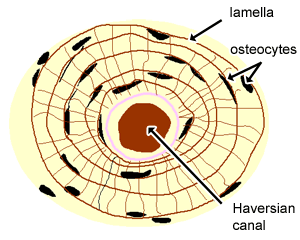
Cartilage, Bone & Ossification: The Histology Guide from www.histology.leeds.ac.uk He was a japanese professor and a quality management innovator of his time. Terms in this set (25). Also called cortical bone, the compact variety usually features a haversian system, or cylindrical unit within the structure. A central tube called a haversian canal typically runs in the same path as the length of the bone, and contains. The osteon consists of a central canal called the osteonic (haversian) canal, which is surrounded by concentric rings (lamellae) of matrix. Your bones are strong enough to support your weight, but light enough to allow movement. Download scientific diagram | structure of compact bone. Cheek bone (zygoma) upper jaw (maxilla).
Cortical bone is compact bone, while cancellous bone is trabecular and spongy bone.
Cortical bone is compact bone, while cancellous bone is trabecular and spongy bone. They protect your delicate internal organs and act as a storehouse for minerals, such as calcium. Also called cortical bone, the compact variety usually features a haversian system, or cylindrical unit within the structure. Hematoma formation, fibrocartilaginous callus formation, bony callus formation, and remodeling. Cheek bone (zygoma) upper jaw (maxilla). We'll go over all the flat bones in your body, from your head to your pelvis. Begin by identifying the concentric rings of lamellar bone that surround a haversian canal. The femur and tibia already bear the weight of most of the body; The osteon consists of a central canal called the osteogenic (haversian) canal, which is surrounded by concentric rings (lamellae) of the matrix. What is the innermost part of the bone called? Compact bone diagram bone cross section diagram file624 diagram of compact bone new. The osteon consists of a central canal called the osteonic (haversian) canal, which is surrounded by concentric rings (lamellae) of matrix. Flat bones are a specific type of bone found throughout your body.
He was a japanese professor and a quality management innovator of his time. A bone is a rigid tissue that constitutes part of the vertebrate skeleton in animals. Compact bone, dense bone in which the bony matrix is solidly filled with organic ground substance and inorganic salts, leaving only tiny spaces that contain the osteocytes, or bone cells. A central tube called a haversian canal typically runs in the same path as the length of the bone, and contains. Compact bone diagram simple diagram system.
Chapter 6: Osseous Tissue and Bone Structure Flashcards | Easy Notecards from www.easynotecards.com Lower jaw (mandible) collar bone. The radius and ulna are two the radius is the bone which is present laterally, which means when your palm is facing upwards, it is the ulna is the medial bone, which is towards the midline of your body. Related online courses on physioplus. Rethinking pain education learn how to teach your patient about their pain powered by physiopedia start course. These bones tend to support weight and help flat bones: Begin by identifying the concentric rings of lamellar bone that surround a haversian canal. These are mostly compacted bone with little marrow and include most of the bones in the limbs. Compact bone consists of outer and inner sheets of lamellar bone (not seen here) and haversian systems, shown here, that run parallel to the long axis of bones.
Sclerostin inhibits bone formation mostly by antagonizing lrp5/6, thus inhibiting wnt signaling.
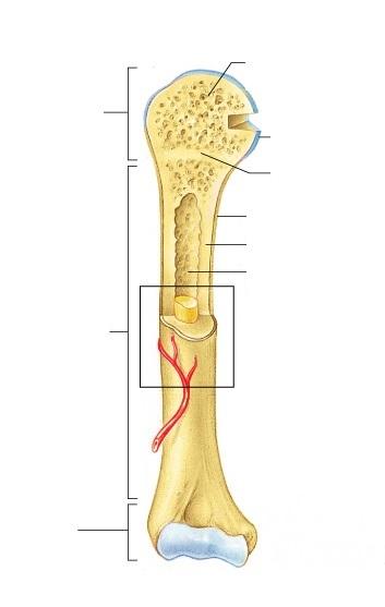
Rethinking pain education learn how to teach your patient about their pain powered by physiopedia start course. There is a printable worksheet available for download here so you can take the quiz with pen and paper. The osteon consists of a central canal called the osteonic (haversian) canal, which is surrounded by concentric rings (lamellae) of matrix. Hematoma formation, fibrocartilaginous callus formation, bony callus formation, and remodeling. Cortical bone is compact bone, while cancellous bone is trabecular and spongy bone. They consist of two outer layers of compact bone and an inner layer of spongy bone. Download scientific diagram | structure of compact bone. Compact bone consists of closely packed osteons or haversian systems. These are mostly compacted bone with little marrow and include most of the bones in the limbs. To resist these stresses, the material should be as far from the neutral axis as diagram showing computed lines of constant stress from the analysis of various transverse sections. Edraw is a new uml diagram and software diagram drawing tool. A diagram of the anatomy of a bone, showing the compact bone. Compact bone consists of closely packed osteons or haversian systems.
Despite appearing dry and lifeless, your bones are a hive of activity. Compact bone becomes more brittle, particularly in the long bones, which is why breaks and fractures in the tibia and femur are things to keep note of when diagnosed with osteoporosis. Have you ever broken a bone? The osteon consists of a central canal called the osteogenic (haversian) canal, which is surrounded by concentric rings (lamellae) of the matrix. Rethinking pain education online course:
Print Exercise 9: Overview of the Skeleton: Classification and Structure of Bones and Cartilages ... from www.easynotecards.com Download scientific diagram | structure of compact bone. They protect your delicate internal organs and act as a storehouse for minerals, such as calcium. Compact bone becomes more brittle, particularly in the long bones, which is why breaks and fractures in the tibia and femur are things to keep note of when diagnosed with osteoporosis. The femur and tibia already bear the weight of most of the body; Compact bone diagram simple diagram system. A diagram of the anatomy of a bone, showing the compact bone. A central tube called a haversian canal typically runs in the same path as the length of the bone, and contains. Cortical bone is compact bone, while cancellous bone is trabecular and spongy bone.
Cheek bone (zygoma) upper jaw (maxilla).
There is a printable worksheet available for download here so you can take the quiz with pen and paper. Lower jaw (mandible) collar bone. Terms in this set (25). Rethinking pain education learn how to teach your patient about their pain powered by physiopedia start course. Rethinking pain education online course: Hematoma formation, fibrocartilaginous callus formation, bony callus formation, and remodeling. Compact bone diagram bone cross section diagram file624 diagram of compact bone new. List the steps in the repair process of a simple fracture. This is an online quiz called structure of compact bone. Cortical bone is compact bone, while cancellous bone is trabecular and spongy bone. These are mostly compacted bone with little marrow and include most of the bones in the limbs. Simple bone diagram barca fontanacountryinn com. The remainder is spongelike cancellous bone.
Rethinking pain education online course: compact bone diagram. We'll go over all the flat bones in your body, from your head to your pelvis.
Share :
Post a Comment
for "Simple Compact Bone Diagram : Compact Bones vs. Spongy Bones: What is The Difference? | Diffzi"


Post a Comment for "Simple Compact Bone Diagram : Compact Bones vs. Spongy Bones: What is The Difference? | Diffzi"